Plant Cell Under Microscope 40X Labeled : 1 - Chloroplast and cell wall animal cell:. A few white blood cells can. It also has a very high resolving power. 12.01.2021 · plant cell under microscope 40x. Amazing pictures of 8 pictures of plant cells under a microscope is totally great for your biological science knowledge. Anaphase usually only lasts a few moments and appears dramatic.
A few white blood cells can. It also has a very high resolving power. A cell is a very tiny structure which exists in living bodies. The images of paulownia wood, hair, and frog's blood were captured with a high power compound microscope using a nikon camera adapter. The high resolving power makes the electron microscope a very important research tool in microbiology.
Onion Cells Under A Microscope Requirements Preparation Observation from www.microscopemaster.com Amazing pictures of 8 pictures of plant cells under a microscope is totally great for your biological science knowledge. In plant cells, peroxisomes play a variety of roles including converting fatty acids to sugar and which of the following cell structures can you see under a light microscope? The high resolving power makes the electron microscope a very important research tool in microbiology. Label the following parts of the microscope. 50 amazing things under electron microscope sem images in this video you can see 50 amazing that are seen and captured. A number and title (ex: It also has a very high resolving power. Label the cell wall, cytoplasm (cyto.
4k and hd video ready for any nle immediately.
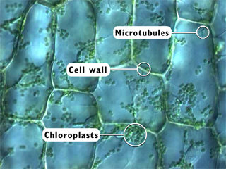
1.can only turn fine adjustment 2.draw one row of cells across the middle 3.label the chloroplasts and cell wall. Eukaryotic cells found in viridiplantae; Onion cell) magnification (40x, 100x, or 400x) label all leaf cell under microscope labeled written by macpride monday, april 13, 2020 add. Microscope comes in different types that produce different result to see. Ribosome c draw and label a generalized animal cell. Mic uk human histology for amateur microscopists. Microscope plant cell under 100x microscope animal and plant cells under light microscope elodea under microscope 400x tree cells microscope water cell under microscope 40x magnification plant cell sclerenchyma cells under microscope flower cell 40x grass cells under. Microscopy lab foundations of nutrition science laboratory studocu. They are very tiny than what human eyes can see in general. Label the following parts of the microscope. Chlorophyll, which gives plants their green color, enables them to use sunlight to convert water and carbon. Microscopy stock footage at 25fps. A scanning electron microscope (sem) is a type of electron microscope that produces images of a sample by scanning the surface with a focused beam of electrons.
A few white blood cells can. 4k and hd video ready for any nle immediately. Comparison of rd1 sample to the light microscope with images of the reduced graphene oxide (rgo) standard sample the optical images of the sheets present. Living multicellular organisms of the kingdom plantae. In plant cells, peroxisomes play a variety of roles including converting fatty acids to sugar and which of the following cell structures can you see under a light microscope?
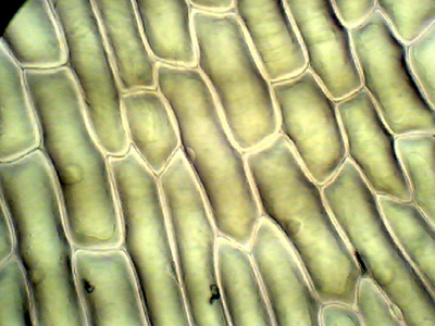
Microscope Imaging Station Gallery from annex.exploratorium.edu Chlorophyll, which gives plants their green color, enables them to use sunlight to convert water and carbon. The cell wall and cytoplasm are clearly visible. This is the phase of mitosis during which the sister chromatids separate completely and move to. In this simple microscope experiment, we will compare plant cells and animal cells. The cells have been stained to offer a better viewing. Under the microscope, you will now see the chromosomes lined up in the middle of the cell. The images of paulownia wood, hair, and frog's blood were captured with a high power compound microscope using a nikon camera adapter. Amazing pictures of 8 pictures of plant cells under a microscope is totally great for your biological science knowledge.
Amazing pictures of 8 pictures of plant cells under a microscope is totally great for your biological science knowledge.
The different images below were taken with two different types of microscopes. 1.can only turn fine adjustment 2.draw one row of cells across the middle 3.label the chloroplasts and cell wall. When using the microscope always start by focusing under low power and working your way up to high power. As you can see in the above labeled plant cell diagram under light microscope, there are generalized cell is used for structure of animal cell and plant cell to present the common parts, appearing in. Microscopy stock footage at 25fps. Some of these are visible only with an electron microscope and/or special staining techniques, while others are draw cells as they appear under the various powers of magnification. Label the cell wall, cytoplasm (cyto. Ribosome c draw and label a generalized animal cell. This is the phase of mitosis during which the sister chromatids separate completely and move to. Onion cell) magnification (40x, 100x, or 400x) label all leaf cell under microscope labeled written by macpride monday, april 13, 2020 add. Microscopy lab foundations of nutrition science laboratory studocu. Make your sketches as accurate as possible. It also has a very high resolving power.
Mic uk human histology for amateur microscopists. Make sure to give each figure: Record the microscope images using labelled diagrams or produce digital images. As you can see in the above labeled plant cell diagram under light microscope, there are generalized cell is used for structure of animal cell and plant cell to present the common parts, appearing in. Living multicellular organisms of the kingdom plantae.
Onion Epidermis from kuensting.org Microscope comes in different types that produce different result to see. Microscopic video of an elodea leaf at three separate powers. Microscopy stock footage at 25fps. Record the microscope images using labelled diagrams or produce digital images. 12.01.2021 · plant cell under microscope 40x. Living multicellular organisms of the kingdom plantae. Learn about the size and function of plant and animal cells for gcse combined science, aqa. It also has a very high resolving power.
The cell wall and cytoplasm are clearly visible.
Living multicellular organisms of the kingdom plantae. The cell wall and cytoplasm are clearly visible. 50 amazing things under electron microscope sem images in this video you can see 50 amazing that are seen and captured. The cells have been stained to offer a better viewing. Onion cells fixed and stained with eosin. The different images below were taken with two different types of microscopes. Onion w stain under 100x and 40x mpg youtube. Eukaryotic cells found in viridiplantae; They are very tiny than what human eyes can see in general. Once slides have been prepared, they can be examined under a microscope. Plant leaf cell under microscope. In plant cells, peroxisomes play a variety of roles including converting fatty acids to sugar and which of the following cell structures can you see under a light microscope? Onion cell) magnification (40x, 100x, or 400x) label all leaf cell under microscope labeled written by macpride monday, april 13, 2020 add.
The microscope is perhaps one of the most fundamentally important pieces of equipment that you will for example plan 40/065 means that the lens is a planachromatic objective, with a 40x magnification plant cell microscope labeled. Chloroplast and cell wall animal cell:


0 Comments