Diagram Of An Animal Cell Under A Microscope - How These 26 Things Look Like Under The Microscope With Diagrams - Cell membrane, nucleus, and cytoplasm with frog red blood cells.
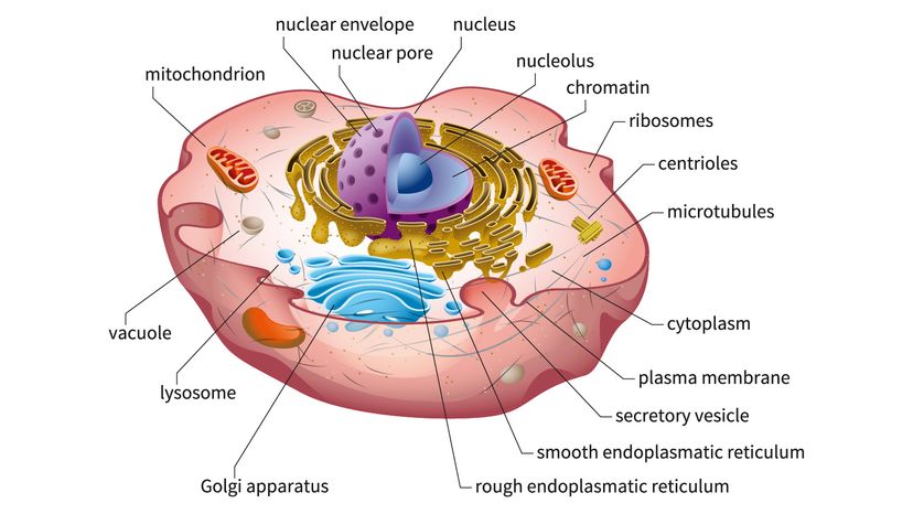
Diagram Of An Animal Cell Under A Microscope - How These 26 Things Look Like Under The Microscope With Diagrams - Cell membrane, nucleus, and cytoplasm with frog red blood cells.. Place the glass slide onto the stage. Cells of plant or animal tissue. The diagram of plant and animal cell structure helps to understand differences and similarities. See how a generalized structure of an animal cell and plant cell look with labeled diagrams. Under the microscope, an animal cell shows many different parts called organelles, that work together to keep the cell functional.
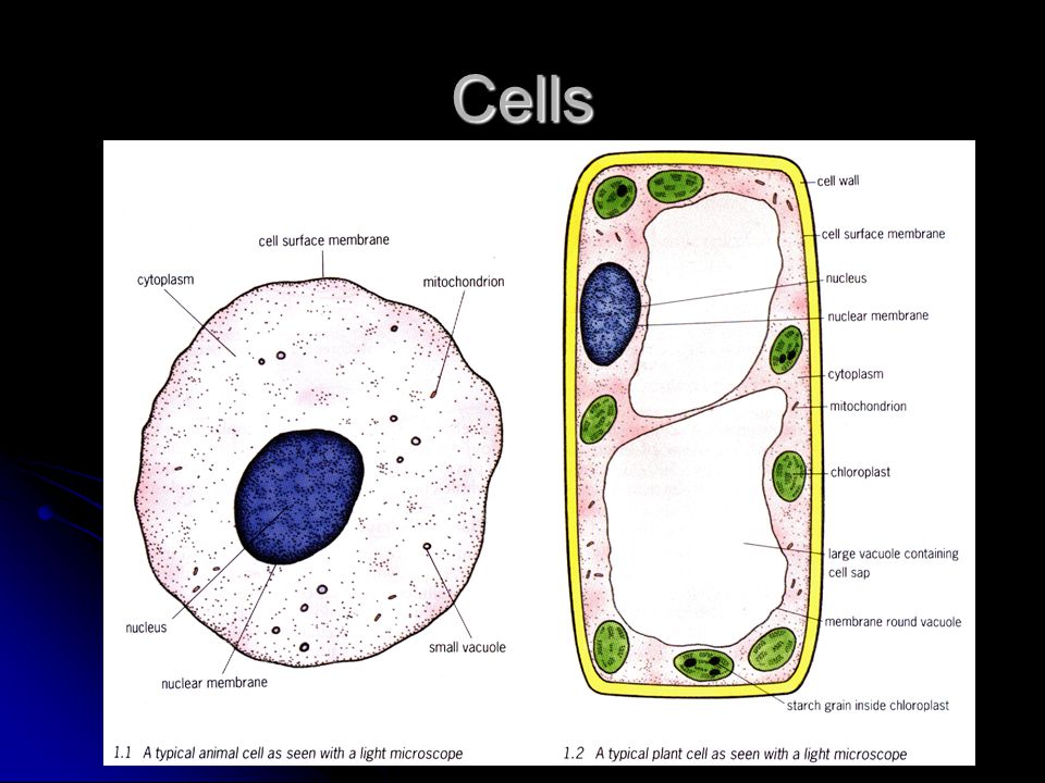
Here's a photo of a plant cell under an electron microscope. Given below is the diagram of a cell as seen under the microscope after having been placed in a solution Light microscopes using visible light and lenses to form a magnified image of the object under investigation e.g. Cheek cells under a microscope. Under a microscope, plant cells from the same source will have a.
Here S How Plant And Animal Cells Are Different Howstuffworks from media.hswstatic.com This is because the stain will color some parts of the cell and not others, allowing them to be clearly. Each cell can live alone sketch your favorite paramecium. Cells under microscope foto sin derechos de autor. Have students draw a diagram of what they see under. • draw a diagram of several blood cells under high power and label their parts. All living organisms are made up of cells. Ultrastructure of a plant cell differences between prokaryotic and eukaryotic cells prokaryotic the cell as whole is in effect, divided up into compartments. Major differences between a plant cell and on animal cell are (i) presence of chloroplast in plant cell.
Under a microscope, plant cells from the same source will have a.
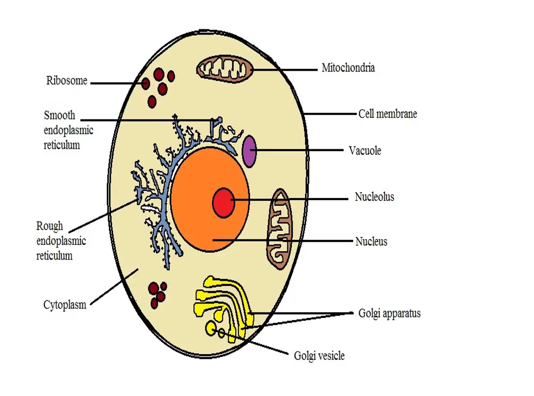
Made the first compound microscope and observed a slice of a cork oak tree. A cell is the structural and functional unit of life. Here's a photo of a plant cell under an electron microscope. Light microscopes using visible light and lenses to form a magnified image of the object under investigation e.g. This is a diagram of a typical plant cell. Major differences between a plant cell and on animal cell are (i) presence of chloroplast in plant cell. (iii) presence of cell wall. This compartmentation is often achieved by membranes so that just as a cell surface. Line diagram of a general animal cell. Cell biology (also called cellular biology or cytology) is the study of cells. • draw a diagram of several blood cells under high power and label their parts. But at the same time it is interpretive. You can specify conditions of storing and accessing cookies in.
Using biological stains such as methylene blue, it's possible to clearly observe and differentiate the different parts of a cell. Light microscopes using visible light and lenses to form a magnified image of the object under investigation e.g. Place a drop of water on your have students use microscopes to observe the shapes of plant and animal cells. We say cells are microscopic because they can only be seen under a microscope. Cheek cells under a microscope.
What Are The Differences Between A Plant Cell And An Animal Cell from www.microscopemaster.com A comparison of plant and animal cells using labelled diagrams and descriptive explanations. Physiology is the study of the functions of the body at the. Light microscopes using visible light and lenses to form a magnified image of the object under investigation e.g. A scale bar has been marked on the drawing, allowing the. Made the first compound microscope and observed a slice of a cork oak tree. The animal cell is made up of several structural organelles enclosed in the plasma membrane, that enable it to function properly, eliciting mechanisms that benefit the host (animal). A cell is a very tiny structure which exists in living bodies. Ppt structure of plant and animal cells under an electron.
A cell is the structural and functional unit of life.
Cells consist of cytoplasm enclosed within a membrane, which contains many biomolecules such as proteins and nucleic acids.2 most plant and animal cells are only visible under a light microscope, with dimensions between 1. Look at the slide under your microscope starting at low power. Examining plant cells under the microscope. Select the lowest power objective lens. A scale bar has been marked on the drawing, allowing the. Removing cellular waste products from the cell. If you examine plant and animal cells under a microscope you will note major structural differences between both. Each cell can live alone sketch your favorite paramecium. Using biological stains such as methylene blue, it's possible to clearly observe and differentiate the different parts of a cell. Cell membrane, nucleus, and cytoplasm with frog red blood cells. • draw a diagram of several blood cells under high power and label their parts. The working together of all cells gives an animal its. All living organisms are made up of cells.
Cheek cells under a microscope. But at the same time it is interpretive. We say cells are microscopic because they can only be seen under a microscope. Ultrastructure of a plant cell differences between prokaryotic and eukaryotic cells prokaryotic the cell as whole is in effect, divided up into compartments. Students will observe cheek cells under a microscope.
Cells Plant Animal Cells Cells How Many Cells Do We Have In Us How Many Cells Do We Have In Us Brain Has Estimated 1 000 000 000 Neurons One Type Ppt Download from images.slideplayer.com This is because the stain will color some parts of the cell and not others, allowing them to be clearly. (ii) presence of large central vacuole in plant cell. This site is using cookies under cookie policy. Cell is a tiny structure and functional unit of a living organism containing various parts known as organelles. Here's a diagram of a plant cell: Be careful pushing it under the clips that the cover slide doesn't move or crack. Light microscopes using visible light and lenses to form a magnified image of the object under investigation e.g. Look near the decaying leaves of the water.
A cell is a very tiny structure which exists in living bodies.
Look at the slide under your microscope starting at low power. Look near the decaying leaves of the water. (at a minimum you should observe: You can specify conditions of storing and accessing cookies in. The most important structures of plant and animal cells are shown in the diagrams below, which provide a clear illustration of how much these cells have in common. We say cells are microscopic because they can only be seen under a microscope. Given below is the diagram of a cell as seen under the microscope after having been placed in a solution To look at a cell close up we need a microscope. Ppt structure of plant and animal cells under an electron. Be careful pushing it under the clips that the cover slide doesn't move or crack. The working together of all cells gives an animal its. Animal cells are the basic unit of life in organisms of the kingdom animalia. The role and function of the plasma membrane;


0 Comments